
Utah MRI Research Center (UMRC)
MRI Facilities
The Utah MRI Research Center (UMRC) provides physics and technical support for MRI users as well as safety training for all individuals associated with research projects.
Scientists and researchers can schedule time on the MRI scanners below at a rate of $500 per hour. Please see our Current Rate Letter for more details.
We are excited to install Utah's first Siemens Freemax MRI scanner! This scanner uses state of the art AI technology to improve image quality using a low field magnet.

A wide-bore, low-field MRI scanner that uses AI technology for improved image quality.
Unique features:
- BioMatrix sensors and software that automatically detect and provide synchronization signals for respiration motion.
- Deep Resolve AI technology that allows for improved image quality and faster scan times.
Configuration:
- 0.55 Tesla – low-field MRI technology
- 0.7 liters of helium with DryCool Technology, zero helium loss
- Bore size - 80cm
- Bio matrix, contour, and breast coils
- Deep Resolve Boost, Gain, and Sharp
Software Version: Syngo XA60 – high computing and reconstruction performance

A high-performance 3 Tesla MRI scanner for human and large animal research.
Configuration:
- Maximum gradient strength - 80mT/m
- Maximum gradient slew rate - 200T/m/s
- Bore diameter- 60cm
- 64 Receive channels
- Dual-channel RF transmitter
Software Version: XA30

A wide-bore, high-performance 3 Tesla MRI scanner for human and large animal research.
Unique features:
- BioMatrix sensors and software that automatically detect and provide synchronization signals for respiration and cardiac motion.
- 4D flow imaging.
Configuration:
- Maximum gradient strength - 60mT/m
- Maximum gradient slew rate - 200T/m/s
- Bore size - 70cm
- 64 receive channels
- Dual-channel RF transmitter
Software Version: XA50

In addition to the vendor-provided receive coils (head/neck coil, Flex coil, etc.), the UCAIR Coil Lab provides custom-made coils dedicated to research, especially for neurovascular imaging and MRI-guided focused ultrasound.


System phantom for T1, T2, Proton Density qMRI standardization

Diffusion Phantom for ADC qMRI Standardization
Research Highlights
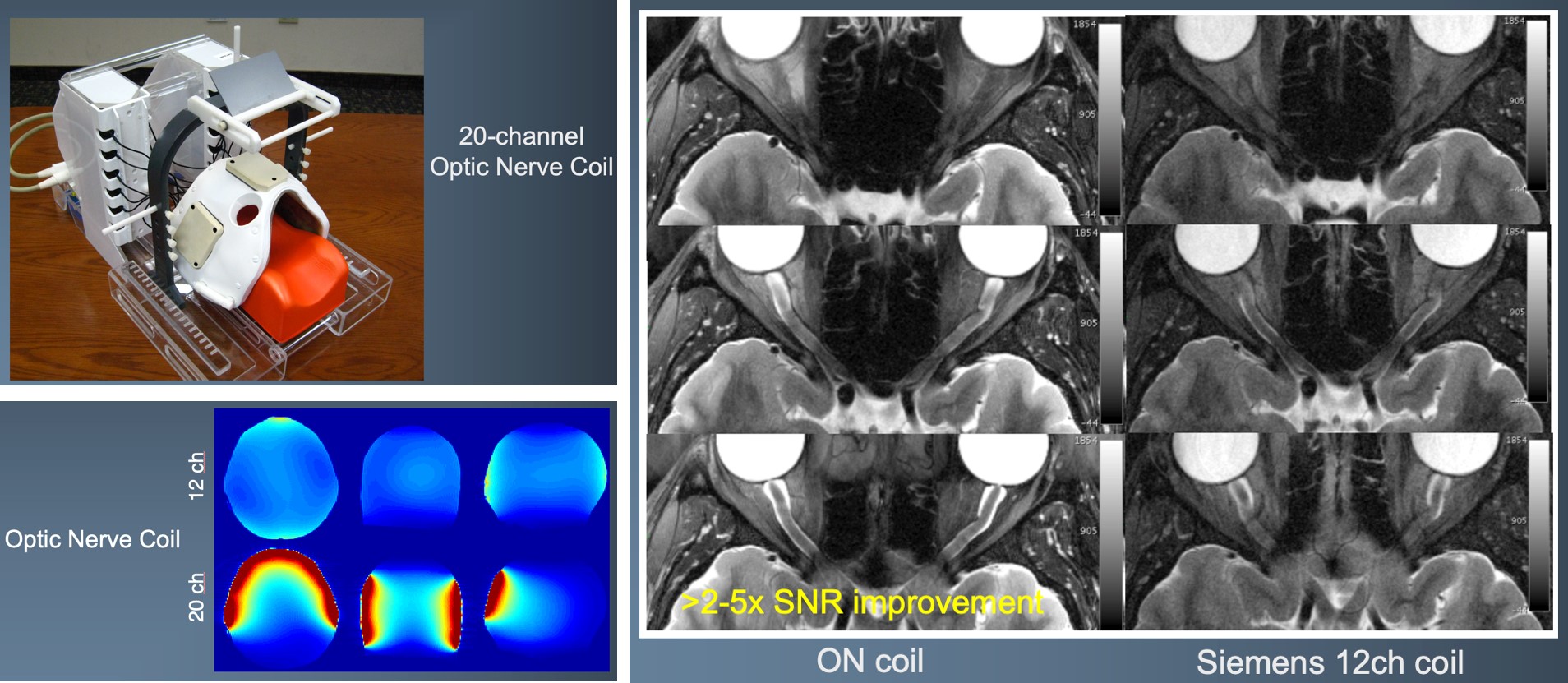
Optic Nerve Visualization
In conjunction with the Custom Coil Lab, researchers developed a much more effective way to visualize the structures associated with the optic nerve.
MR-Guided Focused Ultrasound
The Focused Ultrasound Lab has MR-guided focused ultrasound equipment and expertise that can be applied to a variety of pre-clinical indications. We have numerous protocols that can be utilized for treatment planning, real-time monitoring and treatment assessment.
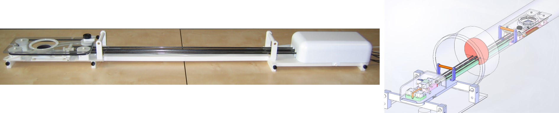
Small animal MR guided focused ultrasound system suitable for mice and rat studies. Other adaptations can be used for small animal neurologic applications. We also have equipment suitable for a variety of large animals.
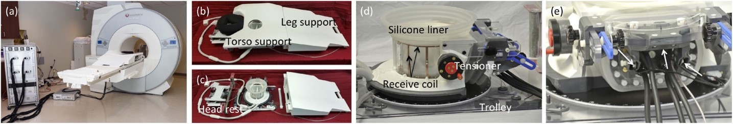
We have also worked with teams to develop MR guided focused ultrasound equipment and protocols for specific clinical applications. Shown is a breast MRgFUS system currently utilized in clinical trials.
MRI-guided high intensity ultrasound to improve drug delivery in spinal cord injury
Donna J. Cross, Allison H. Payne, Henrik Odeen, Yoshimi Anzai, (Radiology)
Candace Floyd, Lonnie Schnieder (PM&R)
Spinal cord injury (SCI) affects thousands of people every year in the USA, and most patients are left with some permanent paralysis. Therapeutic options are limited and only modestly affect outcome. To address this issue, we used magnetic resonance imaging-guided focused ultrasound (MRgFUS) as a non-invasive approach to increase permeability in the blood-spinal cord barrier (BSCB). In this study, we have opened successfully the blood spinal cord barrier in the thoracic region of the normal rat spine using magnetic resonance-guided focused ultrasound combined with microbubbles, and we have measured this quantitatively.
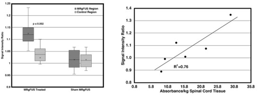
Signal intensity of targeted regions was increased between MRgFUS and sham groups (p=0.003). (b) Evan’s blue tissue absorbance for fixed, excised cord segments correlated to the corresponding region signal intensity on T1w MRI. Cross, Payne et al, 2021 Med Phys.
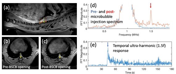
Acoustic emission control for MRgFUS BSCB opening in rats with laminectomy. (a) Post- opening contrast-enhanced sagittal T1w image, arrows indicate sonication locations. Axial CE-T1w image (b) pre- and (c) post-opening with yellow arrow in (a) showing a 14% signal increase at opening site. Acoustic emission frequency spectrum measured during BSCB opening (d) before (blue line) and after (red line) the injection of microbubbles. The ultra-harmonic (1.5 MHz) is readily visible during the microbubble injection (red arrow) allowing for the real-time feedback control of applied MRgFUS pressure at the treatment site.
Contact Us
Need to schedule research time on an MRI at the University of Utah?

Imaging and Neurosciences (INC) Building
729 Arapeen Drive
Salt Lake City, UT 84108-1217